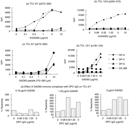
Fig. 2.
Differential presentation by antigen-specific B cell clones, DPA, DPC and DPD of GAD65 to DRB1*0401-restricted T cell lines, measured in a proliferation assay by [3H]-thymidine incorporation. As control, a non-antigen-specific B cell clone, DS389 developed from a DRB1*0401 type I diabetic patient (with no islet cell reactivity) was used. (a) Presentation of the p270–283 epitope of TCL 6/7 by the B cell clones. In greater than five experiments with presentation of GAD65 by DPA and DPD cells, there has been a consistent enhancement of T cell stimulation in comparison to DPC and DS389 cells. (b) Presentation of the p556–575 epitope of TCL 15/3 by the B cell clones. (c) Presentation of the peptide p270–283 by DPA, DPC, DPD and DS389 cells. The experiment with all the four B cell clones was repeated twice with similar results. (d) Presentation of the p106–125 epitope of TCL 15/1 by the B cell clones. The T cell presentation experiments by the autoreactive and DS389 cell lines were repeated three times with similar results. All proliferation assays were conducted in triplicate cultures, but for simplicity the s.e.m. bars are not shown. The symbols of each of the B cell clones are shown in (d). (e) Soluble immune complexes at suboptimal concentrations of GAD65 and purified DPC antibody led to potentiation of presentation at 1·25 and 5·0 µg/ml GAD65 to TCL 6/7 using PBMC as APCs. The proliferative response (average cpm of triplicate cultures) in the presence of the antigen GAD65 and no DPC antibody (100% values) were 597cpm at 0·16 µg/ml GAD65, 1262cpm at 1·25 µg/ml GAD65 and 2092cpm at 5 µg/ml GAD65. The experiment was performed twice with broadly similar results.

