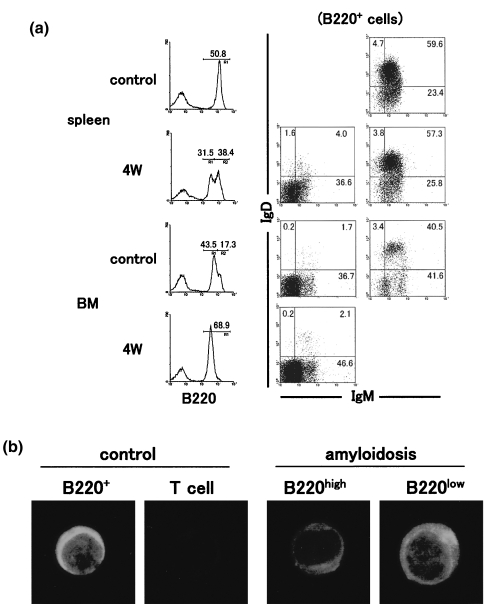
Fig. 4.
Characterization of B220low B cells in the spleen and bone marrow of amyloidosis mice. (a) Three-colour staining for B220, IgM and IgD. (b) Immunofluorescence test of lymphocyte subsets for IgM. After three-colour staining for B220, IgM and IgD, gated analysis was conducted. Numbers in the figure indicate the fluorescence-positive cells in corresponding areas. Lymphocyte subsets were purified from the spleen in amyloidosis mice by a cell sorter and the fixed subsets were stained with FITC-conjugated anti-µ mAb.

