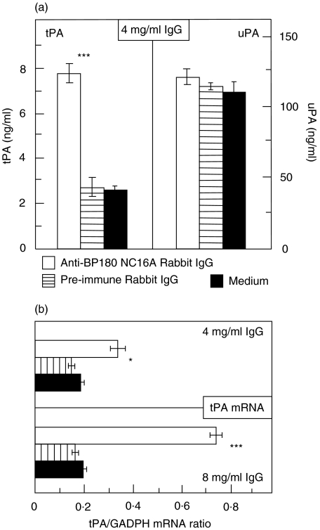
Fig. 5.
Antibodies to BP180 induced both elevated tPA protein and mRNA levels in cultured normal human epidermal keratinocytes (NHEK). Cultured NHEK were treated for a 9 h incubation time with 4 and 8 mg/ml IgG from rabbit R594 (immunized against recombinant human BP180 NC16A; □), with preimmune rabbit IgG ( ), and with medium alone (▪). (a) Culture supernatants were assayed for tPA and uPA reactivity in quadruplicate by ELISA. Bars indicate mean ± SD (ng/ml). (b) tPA mRNA was detected by RT-PCR and expressed as ratio to GAPDH. Asterisks indicate statistical significant difference between R594 IgG- and preimmune IgG-treated cells (*P < 0·05, ***P < 0·001). This pattern is representative of the pattern seen in 2 separate experiments with keratinocytes from different donors.

