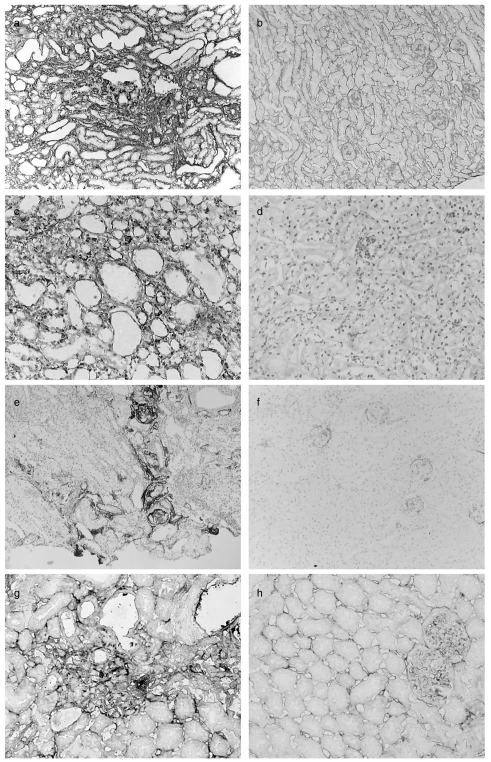
Fig. 4.
Immunohistochemical analysis of kidneys adriamycin-treated mice (a, c, e, g) and control mice (b, d, f, h). There is increased staining for Col IV diffusely throughout the cortex in a peritubular distribution, but particularly in areas of more severe damage (a). Staining of tissue from saline-treated mice is shown as control (b). Staining for HSP47 is increased throughout the cortex of adriamycin-treated mice (c) compared to saline-treated mice (d). C3 (e) and C9 (g) deposition was seen in areas of most severe damage in adriamycin-treated mice. In control mice, very weak staining for C3 was observed only in the glomeruli. Magnification (a, b × 150; c-f × 200; g, h × 250).

