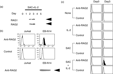
Fig. 2.
RAG protein expression in normal B cells. a. Normal circulating B cells were stimulated with SAC + IL-2, and RAG protein expression was analysed by immunoblotting. The B cells expressed RAG1 and RAG2 when stimulated with SAC + IL-2 for 2–3 days. Equal amounts of the cellular proteins were analysed. Arrows indicate RAG1 (105 kD) and RAG2 (56 kD). The result shown is a representative of 8 independent experiments. b. Intracytoplasmic RAG2 expression of authentic RAG+ (EB-N14; an EB virus transformed cell line) and RAG- (Jurkat T cell line) cells was analysed with flow cytometry and immunoblotting analysis. An arrowhead indicates RAG2 (56 kD). c. Normal B cells were stimulated with either IL-2, SAC or a combination of SAC + IL-2 and intracytoplasmic RAG2 expression was studied by a flow cytometer. The B cells expressed RAG2 protein only when stimulated with SAC + IL-2. The result shown is a representative of 4 independent experiments.

