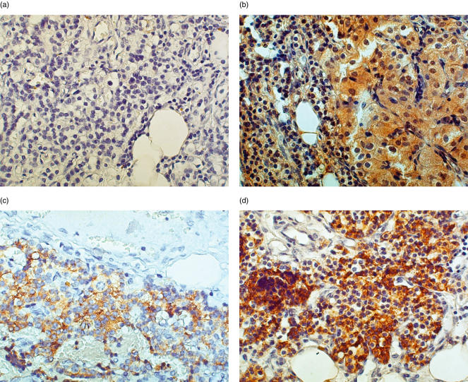
Fig. 2.
A representative sample of parathyroid tumour (ID 4, Table 1) stained for IL-6, PTH and chromogranin-A. Tissue from a patient with familial primary adenoma was embedded in paraffin and stained by immunohistochemistry as described in Materials and Methods. (a) Negative control, 400×; (b)IL-6 staining, 400×; (c) PTH staining, 400×; (d) Chromogranin-A staining, 400×.

