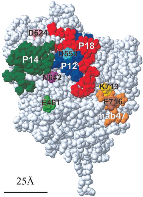
Fig. 2.
Space filling representation of the modelled MPO-like domain of TPO [7]), showing the location of the single amino acids replacements and the peptides used in this study. Peptide P14 (green) principally defines IDR-B [7]. P12 (blue) and P18 (red), together with P14, define a region that encompasses both IDR-A and -B domains. The sequence comprising the epitope for mab 47 (residues 713–721), is shown in orange, including the single amino acid replacement residues within this region, K713 (yellow) and E716 (brown) highlighted. Peptide P43 (residues 702–721) includes this sequence. Also shown are the other single amino acid replacements D624 (pink), D553 (light blue), N642 (purple) and E461 (light green). The bar (25 Å long) represents the approximate diameter of an antibody combining site.

