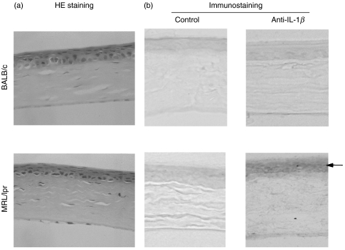
Fig. 3.
Histological and immunohistochemical examinations of corneas from MRL/lpr and BALB/c mice. (a) Representative histological pictures of corneas showing no significant differences between the two strains. (H&E staining, × 400) (b) Representative pictures of IL-1β expression in corneas: left; without specific antibodies, right; stained with anti-IL-1β antibodies. IL-1β is specifically localized in the corneal epithelial layer and was observed only in an MRL/lpr strain. (×400)

