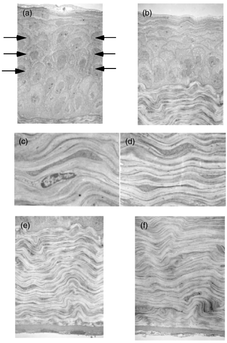
Fig. 4.
Transmission electron microscopy of the corneas of BALB/c and MRL/lpr mice (Original magnification × 2700). No inflammatory cells were seen in any layers of the corneas of both strains. The wing cells in (b) the corneal epithelia of MRL/lpr mice were decreased compared with (a) those of BALB/c (arrows). No differences were seen in the structure or dimensions of the corneal stroma or endothelial cells between MRL/lpr (c,e) and BALB/c (d,f) mice.

