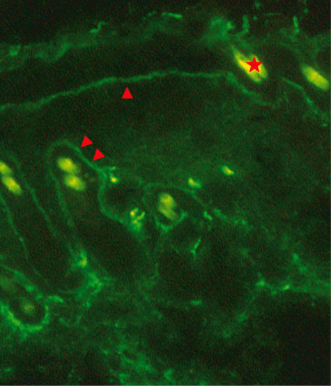
Fig. 4.
Fluorescence photograph of IgG deposition. IgG deposition is observed at the dermo–epidermal junction (arrowhead) and at the basement membrane zone of the hair follicle (double arrowheads). Autofluorescence of hair is seen, one of which is indicated by an asterisk (original magnification × 100).

