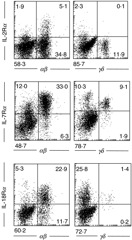
Fig. 3.
Flow-cytometric analysis of IL-2 receptor α, IL-7 receptor α and IL-18 receptor α with freshly isolated IELs. Cells were stained with PE-conjugated anti-IL-2Rα, IL-7Rα or IL-18Rα MoAb and FITC-conjugated antihuman TCRαβ or γδ MoAb. After washing, a minimum of 10 000 cells was analysed by FACScan. The results are representative of three similar experiments.

