Fig. 5.
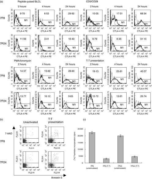
T cells rendered anergic express high levels of CTLA-4. 7P8 and 7P24 cells were stimulated with either anti-CD3 and anti-CD28 antibodies, PMA and ionomycin, peptide pulsed BLCLs, or by T:T presentation of peptide. (a) Cell surface CTLA-4 expression in response to the various stimuli was measured after two, four, and 24 hours. Isotype controls are shown as thin lines, and anti-CTLA-4PE mAb as thick lines. (b) Parallel cultures were set up which were stimulated for 24 hours, after which time the cells double stained with the vital dyes 7AAD and annexin 5, and counted using tru-count beads. (c) Proliferation of unstimulated cells or cells stimulated by T:T presentation, in response to subsequent challenge by BLCL presenting specific peptide, was then investigated. Recovered cells were cocultured with peptide-pulsed BLCL, at concentrations of 5 × 10³ and 3 × 10⁴, respectively. 3H thymidine incorporation was used as a measure of proliferation.

