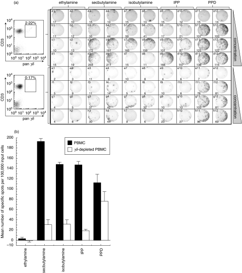
Fig. 1.
γδ T cells respond to alkylphosphate, alkylamine and PPD antigens directly ex vivo. (a) Demonstration of γδ T cell activation by direct ex vivo IFNγ ELISpot assay. The top half of the plate was set up with 100 000 PBMC/well and the bottom half with 100 000 γδ-depleted PBMC per well. FACS analysis with pan-γδ and anti-CD3 antibody (left) showed that γδ-depletion was >92% successful. Cells were activated with ethylamine in columns 1 & 2, secbutylamine in columns 3 & 4, isobutylamine in columns 7 & 8, IPP in columns 9 & 10 and PPD in columns 11 & 12. Two replicates were performed for each experimental condition. Rows a and e are control rows without added antigen. Alkylamine antigens were added at 1 mm in rows b & f, 10 mm in rows c & g and 50 mm in rows d & h. IPP was added at 10 µm in rows b & f, 100 µm in rows c & g and 1 mm in rows d & h. PPD was added at 5 µg/ml in rows b & f, 10 µg/ml in rows c & g and 20 µg/ml in rows d & h. The ELISpot photograph and number of spots (bottom of each panel) were generated mechanically using an ELISpot Reader System ELR02 (Autoimmun Diagnostika; Strassberg). (b) Number of specific spots (sample minus background) per 100 000 PBMC (▪) and γδ-depleted PBMC (□) induced by specified antigens. Bars represent spots induced by 50 mm ethylamine, 50 mm secbutylamine, 10 mm isobutylamine (50 mm isobutylamine was toxic to cells), 1 mm IPP and 20 µg/ml PPD. Error bars show the standard deviation from the mean of two replicate wells.

