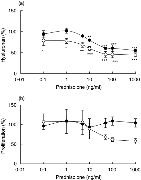
Fig. 2.
Hyaluronan content in supernatants of confluent fibroblasts exposed to different concentrations of prednisolone for 48 h (a) and proliferation, measured as [3H]-thymidine incorporation, of confluent fibroblasts exposed to different concentrations of prednisolone for 48 h (b). The fibroblasts were isolated from cardiac allografts undergoing rejection (•) or from normal heart tissue (○). The results are mean values ± s.e.m. of three or four different experiments where every experiment consisted of triplicates. *P < 0·05, **P < 0·01 and ***P < 0·001 compared with hyaluronan content or proliferation of fibroblasts incubated in the absence of prednisolone.

