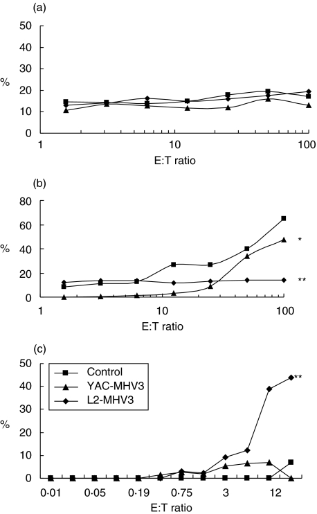
Fig. 3.
Determination of the percentage of cytotoxic activity of the splenic (a), myeloid (b) and intrahepatic (c) NK1·1+ cells from mock-infected (▪), L2-MHV3 (⋄) and YAC-MHV3 (▴) -infected C57BL/6 mice at 72 h p.i. Cytotoxicity of lymphoid cells from liver, spleen and bone marrow against YAC-1 target cells was evaluated by release of LDH activity in the supernatant. The optical density was recorded using an ELISA reader (490 nm filter). The experiments were conducted in triplicate. *P < 0·05; **P < 0·001.

