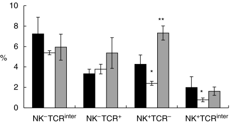
Fig. 5.
Percentages of NK1·1+TCR−, NK1·1+TCRinter, NK−TCRinter and NK−TCRhigh cells in in vitro L2-MHV3- and YAC-MHV3-infected intrahepatic lymphoid cells stimulated with IL-15. Intrahepatic lymphocytes were incubated with 20 ng/ml of rmIL-15 for 7 days, then infected with 0·1–1 m.o.i. mock-infected (▪), L2-MHV3- (□) and YAC-MHV3- ( ) infected for 24 h and immunolabelled with anti-NK1·1+PE and antiαβ-TCR MoAbs. Lymphoid cells were gated according to FSC/SSC parameters obtained on a FACScan flow cytometer and the percentages were evaluated based on a total of 10 000 events recorded. Results are representative of three experiments. *P < 0·05; **P < 0·01.

