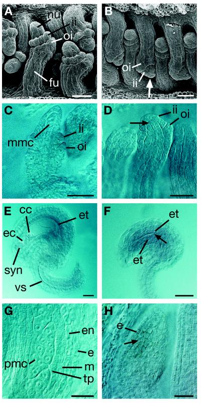
Figure 1.
Summary of the nzz mutant phenotypes. (A and B) Scanning electron micrographs. (C–H). Optical sections through whole-mount tissue. (A, C, E, and G) Wild type. (B, F, and H) nzz-2. (D) nzz-1. Stages: (A–D) ovule stage 2-III, (E and F) ovule stage 3-VI, (G and H) floral stage 7. (A and C) The nucellus is prominent and the two integuments are initiated. (B and D) Note the reduced distal tip region (arrow) and compare with A and C. (E) A fully developed embryo sac and fully developed integuments are seen. (F) There is a prominent absence of a nucellus and an embryo sac at this stage (arrow). The integuments are reduced; however, an endothelium still can be recognized. (G) An anther at a premeiotic stage. The PMCs and the descendants of the primary parietal layer are clearly recognizable. (H) Only a mass of uniform cells is detectable in this anther (arrow). cc, central cell; e, epidermis; ec, egg cell; en, endothecium; et, endothelium, fu, funiculus; ii, inner integument; m, middle layer; mmc, megaspore mother cell; nu, nucellus; oi, outer integument; pmc, pollen mother cell; syn, synergid; tp, tapetum; vs, vascular strand. (Scale bars, 20 μm.)

