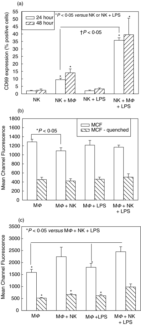
Fig. 1.
Splenic NK cell activation at 24 and 48 h (a) and peritoneal macrophage (MΦ) phagocytosis at 24 h (b) and 48 h (c) in single cell-type and mixed-cell cocultures from wild type C57BL/6 mice. Isolated splenic NK cells were cultured alone and with MΦ with and without LPS. CD69 expression was determined after 24 and 48 h culture, and levels were significantly increased in cocultures with MΦ (NK + MΦ, NK + MΦ+ LPS) (a). MΦ were similarly cultured alone or with NK cells, with and without LPS, and levels of phagocytosis of fluorescent-labeled E. coli determined at 24 h and 48 h. No differences were seen in phagocytosis after 24 h (b). Phagocytosis mean channel fluorescence (MCF) was significantly increased in cocultures of MΦ+NK cells +LPS at 48 h compared with other groups (c). n = 6 per experimental group and data are representative of separate repeated experiments. Data presented as mean ± s.e.m. * or †P < 0.05, Kruskal–Wallis (CD69 percentages), anova (MCF).

