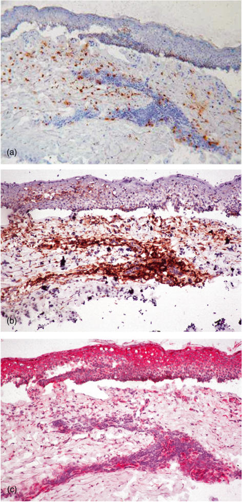
Fig. 4.
Immunohistochemical staining of EG2+ eosinophils (a), CD4+ lymphocytes (b) and IL-16 (c) in serial cryostat sections from a representative sample of bullous pemphigoid (BP) lesional skin, showing immunoreactive IL-16 in both dermal CD4+ infiltrate and epidermal keratinocytes (HRP two-steps amplified method, DAB and AEC chromogen substrates, haematoxylin counterstaining, ×250).

