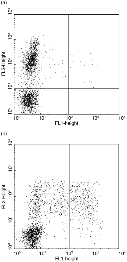
Fig. 1.
PBMC NK cells produce IFN-γ in response to CT EBs. Staining PBMC for expression of CD94-PE (y-axis) and intracellular IFN-γ-FITC (x-axis) after 20·5 h culture in: (a) medium alone, or (b) with CT EBs (10 organisms per cell). Golgi stop was added during the last 6 h of culture. IFN-γ+ CD94+ (NK) cells are readily visible in panel (b) (17%, top R quadrant; population gated on viable CD3- cells).

