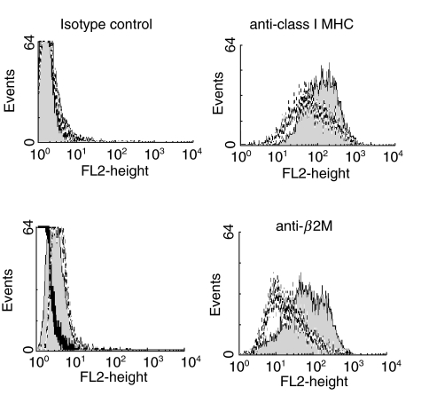
Fig. 5.
CT infection decreases MHC class I antigen expression by epithelial cells. Expression of class I HLA antigens as detected by w6/32 (upper panel and a MoAb against β2m (lower panel) by CT-infected SiHa cells (dotted lines) or mock-infected SiHa cells (filled histograms). Staining with isotype match controls for each MoAb is also shown (left panels). Dead cells, which were more abundant in the CT-infected population, were excluded from analysis by gating on forward scatter.

