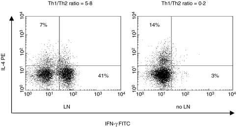
Fig. 5.
Flow-cytometry distribution of circulating Th1 (IFNγ+) and Th2 (IL-4+) in SLE patients divided in relation to the presence of nephritis. The analysis revealed an expansion of the IFNγ+ cell population in LN patients with respect to their control. Unstained cells served to adjust for autofluorescence and arrange quadrants. Percent of positive cells and relative Th1/Th2 ratios are indicated. The results are representative each of 5 patients.

