Fig. 3.
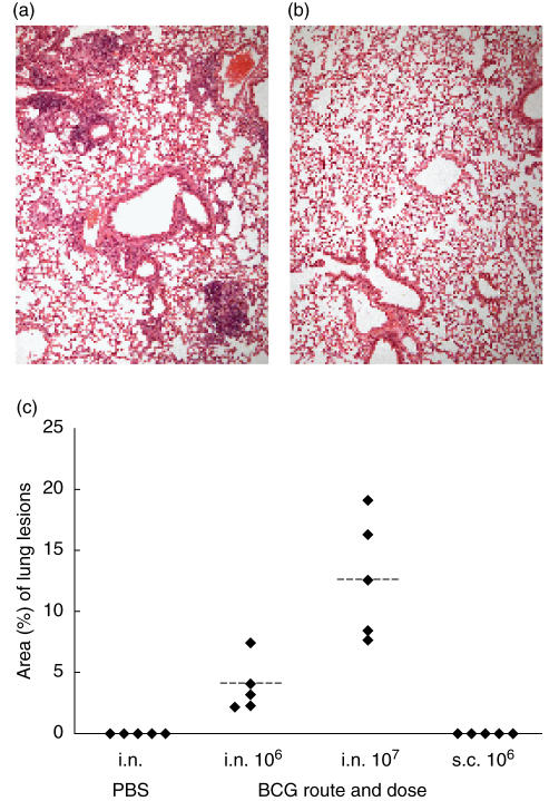
Morphometric analysis of granulomatous infiltration of the lungs. Representative haematoxylin and eosin stained sections of lungs, harvested 12 weeks after intranasal delivery of 107 CFU organisms (a) or phosphate buffered saline (PBS) (b). Lungs from BALB/cJCit mice given 106 CFU BCG subcutaneous were indistinguishable from the intranasal PBS inoculated controls. (c) Individual values of the relative proportion of lung areas with granulomatous lesions from five mice per group after intranasal (i.n.) or subcutaneous (s.c.) BCG infection and intranasal PBS inoculated controls. Dotted lines = mean values.

