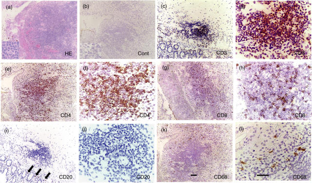
Fig. 1.
Infiltration of mononuclear cells to intestinal lesions of BD. (a) An intestinal ulcer of BD was infiltrated by inflammatory cells; a vast majority were mononuclear cells. A very small number of neutrophils also infiltrated to the site. Inset, higher magnification. (b) Result of staining with control antibody, mouse IgG, is shown. (c) Almost all the mononuclear cells were found to be CD3+ T cells. (d) Higher magnification of (c). (e) A majority of the infiltrated cells to the intestinal ulcer were CD4+ cells. (f) Higher magnification of (e). (g) CD8+ cells constituted a minor population infiltrating to the lesions. (h) Higher magnification of (g). (i) A few CD20+ B cells were found in the lesions (arrows). (j) Higher magnification of (i). (k) CD68+ macrophages resided mainly on the outer side of the aggregate. (l) Higher magnification of (k). Scale bars represent 100 µm for (a) CEGIK and 50 µm for (b) DFHJL. Results of a representative case of BD5 are shown.

