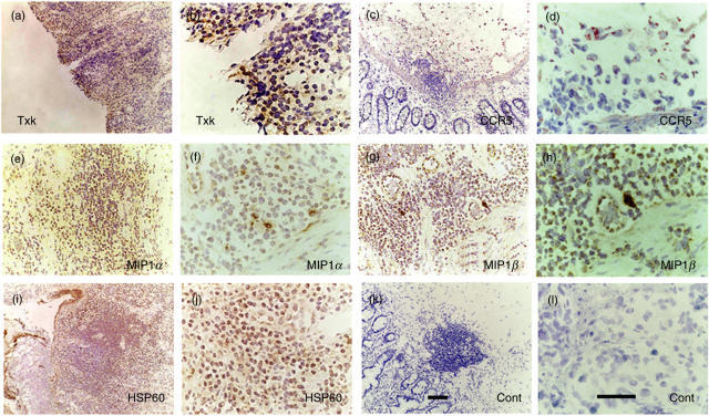
Fig. 3.
Expression of Txk, CCR5, MIP1α, MIP1β and HSP60 in the intestinal ulcer of BD. (a) Txk was expressed in the lymphocytes accumulating to the lesions. (b) Higher magnification of (a). (c) CCR5 was expressed in the mononuclear cell aggregate. (d) Higher magnification of (c). (e) MIP1α was expressed in mononuclear cells. (f) Higher magnification of (e). (g) MIP1β was expressed in mononuclear cells. (h) Higher magnification of (g). (i) HSP60 expressing mononuclear cells located within and outside of the cell aggregates. (j) Higher magnification of (i). (k) Result of a control antibody, mouse IgG, is shown. (l) Higher magnification of (k). Scale bars represented 100 µm for (a) CEGIK and 50 µm for (b) DFHJL. Results of a representative case of BD5 are shown.

