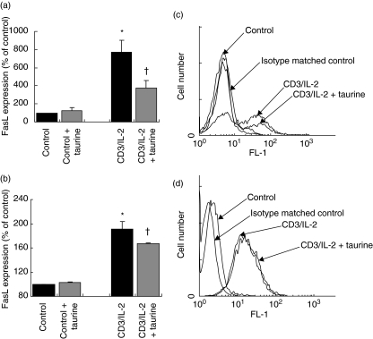
Fig. 2.
Taurine reduces CD3/IL-2-induced FasL expression on T cells. (a) Jurkat cells were stimulated with anti-CD3 MoAb (5 µg/ml) and IL-2 (500 U/ml) for 18 h, and then re-stimulated with IL-2 (500 U/ml) only for an additional 18 h. FasL expression was measured by flow cytometry. Stimulation of Jurkat cells with CD3/IL-2 up-regulated FasL expression relative to unstimulated control (*P < 0·05 versus control). Preloading with taurine (40 m m for 64 h prior to CD3/IL-2 stimulation) significantly reduced CD3/IL-2-induced FasL expression on Jurkat cells (†P < 0·01 versus CD3/IL-2). Data are expressed as the mean ± s.e.m. (n = 5). Statistical analysis was performed by one-way anova with LSD post hoc correction. (b) CD4+ T cells freshly isolated from peripheral blood of male volunteers were stimulated with anti-CD3 MoAb (5 µg/ml) and IL-2 (500 U/ml) for 18 h, and then re-stimulated with IL-2 (500 U/ml) only for an additional 18 h. FasL expression was measured by flow cytometry. Stimulation with CD3/IL-2 significantly up-regulated expression of FasL on freshly isolated CD4+ peripheral T cells compared to unstimulated control (*P < 0·05 versus control). Preloading the cells with taurine (40 m m for 64 h prior to CD3/IL-2 stimulation) significantly down-regulated CD3/IL-2-induced FasL expression on peripheral CD4+ lymphocytes (†P < 0·05 versus CD3/IL-2). Data are expressed as the mean ± s.e.m. (n = 3). Statistical analysis was carried out by one-way anova with LSD post hoc correction. (c) Flow cytometry histogram of Jurkat FasL expression. Binding of anti-FasL antibody is indicated by the arrows. There is no antibody binding to control cells as resting Jurkat cells do not express FasL. (d) Flow cytometry histogram of Jurkat IL-2r expression. Binding of anti-IL-2r antibody is indicated by the arrows. Statistical analysis was performed by one-way anova with LSD post hoc correction.

