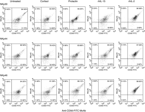
Fig. 2.
Expression of NKp30, NKp44 and NKp46 on peripheral blood NK cells from healthy human donors. Freshly purified peripheral blood NK cells were cultured for 16 h with or without prolactin, cortisol, rhIL-2 or rhIL-15. They were then analysed by direct two-colour immunofluorescence and FACS analysis with PE-conjugated MoAbs against NKp30, NKp44 and NKp46 in combination with FITC-conjugated anti-CD56 MoAb. The dot plots were divided into quadrants representing unstained cells (lower left), cells with only red fluorescence (upper left), cells with red and green fluorescence (upper right) and cells with only green fluorescence (lower right).

