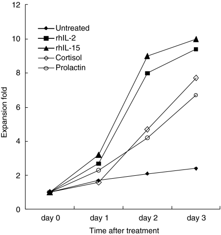
Fig. 4.
Growth of NK cells after exposure to prolactin, cortisol, rhIL-2 or rhIL-15. Peripheral blood NK cells (5 × 10⁶) were cultured at 37°C in a 5% CO2 incubator. Cells were harvested and counted at the indicated times. Cell viabilities were determined by the trypan blue exclusion test and were >95% for all preparations. All time-points represent means of six independent experiments.

