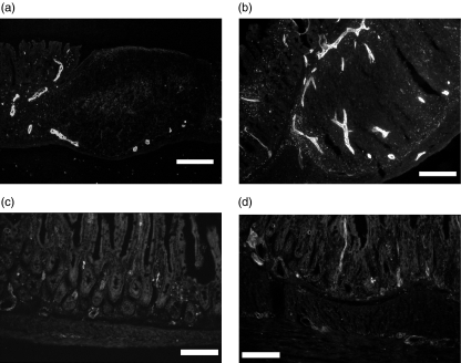
Fig. 1.
Immunohistochemical study of MAdCAM-1 and VCAM-1 expression in the intestinal mucosa. (a) MAdCAM-1 expression of AKR/J mice on the vessel near Peyer's patches (b) MAdCAM-1 expression of SAMP1/Yit mice demonstrating the increased MAdCAM-1 expression (c) VCAM-1 expression of AKR/J mice was weak in the lamina propria. (d) VCAM-1 positive vessels of SAMP-1/Yit mice were increased mainly in the submucosal area compared with those in AKR/J mice. Bar: 100 µm (objective lens × 20).

