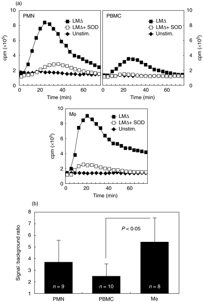
Fig. 1.
LCL of PMN, PBMC and Mφ in response to LMΔ. Addition of superoxide dismutase to LMΔ-stimulated cells (LMΔ + SOD) abrogates the CL signal. Mean temporal traces of two from representative experiments (a) and average signal-to-background ratios of LMΔ-stimulated versus unstimulated cells integrated over the whole experiment (b) are shown. P-values indicate levels of statistical significance of difference.LCL of PMN, PBMC and Mφ by viable and heat-killed LM. Mean temporal traces of two from a representative experiment (a) and mean signal-to-background ratios of viable LM-stimulated versus LMΔ-stimulated cells integrated over the whole experiment (b) are shown for one representative experiment.

