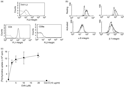
Fig. 1.
Expression of surface antigens on intraepithelial lymphocyte (IEL) cell line established from ovalbumin (OVA)23-3 transgenic mice; 2 × 10⁵ lymphocytes were first incubated with antimouse monoclonal antibodies against Thy1·2 (30-H12), pan T cell receptor (TCR) β chain (H57-597), CD4 (YTS191·1.2), CD8a (53.6.7), αE-intergin (M290), β7-integrin (Fib27), l-selectin (MEL-14) and CD11a (M17/4). They were then incubated with 1 ml of fluorescein isothiocyanate (FITC)-labelled antirat IgG and antihamster IgG. Flow cytometric analysis was performed using FACSort (Becton Dickinson). Data on viable cells, as determined by forward light-scatter intensity, were obtained using consort software. Representative data from at least four individual measurements are shown. (a) Thy 1·2, CD4 and CD8α expression on resting IEL cell line (b); αE and β7 expression on resting and activated IEL cell line. For IEL cell line activation, cells were stimulated by a specific antigen, OVA (20 µm), for 20 h. (c) Antigen-specific and mitogenic proliferation of IEL cell line. Uptake of [3H]-thymidine was examined as described in Materials and methods. Proliferation of IEL cell line in response to different concentrations of ovalbumin (OVA, 1–20 µm) and concanavalin A (ConA, 10 µg/ml) was determined. Values are means ± s.d. from six experiments. *P < 0·05 versus OVA 0 µm.

