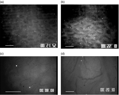
Fig. 2.
Representative images of the distribution of a carboxyfluorescein diacetate succinimidyl ester (CFSE)-labelled resting intraepithelial lymphocyte (IEL) cell line in control mice (a) and in ovalbumin (OVA)-fed mice (b) Following adherance to the microvessels of a villus tip of the ileal mucosa at 20 min after infusion ( × 10). Bar represents 100 µm. (c) Higher magnification image of labelled IEL cell line in control mice adhered to arcade microvessels of villus tips ( × 20). Bar represents 100 µm. (d) Observation of CFSE-labelled resting IEL cell line postcapillary venules of Peyer's patches 20 min after infusion. There were few sticking IELs in this area ( × 10).

