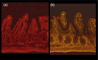
Fig. 4.
Representative pictures of simultaneous observation of carboxyfluorescein diacetate succinimidyl ester (CFSE)-labelled intraepithelial lymphocyte (IEL) (green) and factor VIII-positive (a) or CD34-positive (b) microvessels (red fluorescence) in small intestinal villi as determined by immunohistochemistry. The lysine–paraformaldehyde (PLP)-fixed sections 40 min after IEL infusion were observed (× 100).

