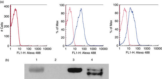
Fig. 2.
Confirmation that erythrocytes (E) from the transgenic mouse expressed human complement receptor 1 (CR1) antigen of appropriate molecular size. (a) human E (hE), transgenic mouse E (mE) and mE + CR1 were stained with Alexa 488 labelled anti-CR1 MoAb 7G9 and analysed by flow cytometry. CR1 antigen was expressed on the surface of the E from the transgenic mouse (centre panel), but not on E from a wild-type mouse (left panel). hE (right panel), which were used as a positive control, expressed about 50% as much CR1 on the mE + CR1. Blue histogram, anti-CR1 mAb; red histogram, isotype control mAb. (b) Immunoblotting for human CR1. Confirmation that transgenic mouse E (mE + CR1) express CR1 protein of appropriate size. Non-reduced lysates of wild-type mE, mE + CR1 and hE were resolved on a 4–12% sodium dodecyl sulphate-polyacrylamide gel electrophoresis (SDS-PAGE) gel and the proteins transferred to nitrocellulose for development with anti-CR1 MoAb YZ-1 as described in Methods. Lane 1, prestained myosin 207 kDa MW marker; lane 2, wild-type mE; lane 3, mE + CR1; lane 4, hE from a donor with CR1*1 and CR1*3 alleles of CR1.

