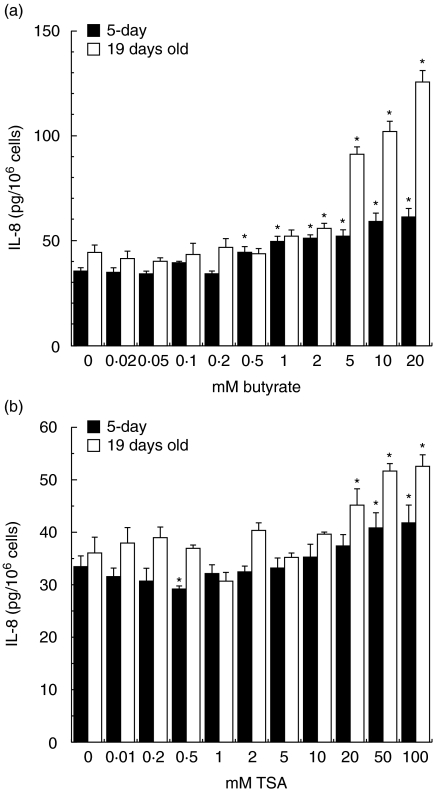
Fig. 7.
Interleukin (IL)-8 secretion in crypt-like (a) and villus-like (b) Caco-2 cells after incubation with butyrate or trichostatin A. The Caco-2 cells were exposed to physiological butyrate concentrations (0–20 m M) and comparative concentrations of trichostatin A (TSA) (0–100 n M) for 24 h. Culture medium was collected. IL-8 secretion is expressed as pg IL-8/106 cells. The IL-8 secretion was determined using two cell passages and triplicate cultures per passage. Significant differences (*P < 0·05) between the levels of IL-8 for cells exposed to butyrate or TSA and control Caco-2 cells are indicated. Caco-2 cells exposed to butyrate (a) and TSA (b).

