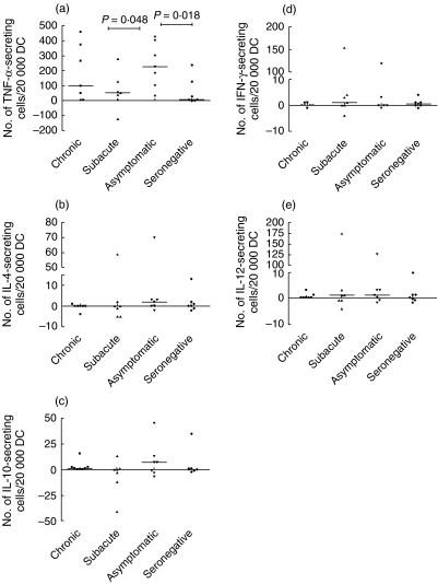
Fig. 2.
The median number of live Borrelia garinii-specific (a) tumour necrosis factor (TNF)-α-, (b) interleukin (IL)-4-, (c) IL-10-, (d) interferon (IFN)-γ- and (e) IL-12p70-secreting cells per 20 000 in vitro differentiated dendritic cells (DCs) from patients with a history of different clinical outcomes of Lyme borreliosis (LB): chronic (including one patient with acrodermatitis chronicum atrophicans (ACA) and subacute neuroborreliosis, asymptomatic seropositive individuals and seronegative, healthy controls. The borrelia-specific cytokine secretion was obtained by subtracting the number of spots in unstimulated wells from the number in the borrelia-stimulated wells. P-values (Mann—Whitney U-test) from comparison between patient groups are shown. Note the different scales on the y-axes.

