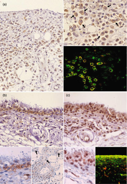
Fig. 2.
Representative immunostaining of chemokines and their receptors in early rheumatoid arthritis (RA) synovium. (a) CXCR3+ cells are localized mainly in the synovial sublining region. These cells are including plasma cells (right, upper) (arrows), and are mostly positive for the plasma cell marker CD138 (right, lower; green, CXCR3, red; CD138 and yellow, double positive) (case 3). (b) Mig/CXCL9, a ligand for CXCR3, is stained along the synovial sublining region, but not the intimal lining layer. The positive cells seem synovial type B cells [inset, left: a higher magnification of (b)]. Mig/CXCL9 is also positive in the perivascular cells and endothelial cells (inset, right) (arrows) (case 4). (c) MEC/CCL28, a ligand for CCR10, is positive mainly in the intimal lining cells showing a palisading structure, corresponding to synovial type A cells [inset, left: a higher magnification of (c)]. MEC/CCL28 co-localizes mostly with CD68 (inset, right) (green, CXCR3; red, CD138 and yellow, double positive) (case 4).

