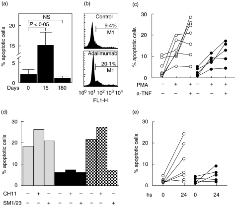
Fig. 4.
Induction of apoptosis of PBMNC by anti-TNF-α agents. (a) Fresh isolated PBMNC from eight RA patients were fixed, stained by TUNEL technique, and analysed by flow cytometry. Data correspond the arithmetic mean and SE of eight RA patients at the indicated times of Adalimumab therapy. (b) PBMNC from healthy individuals were cultured in the presence or not of 10 µg/ml Adalimumab for 24 h, and then apoptotic cells were quantified by TUNEL and flow cytometry analysis. Data of an experiment out of six performed are shown. The percent of apopotic cells is indicated. (c) PBMNC from 12 RA patients were preactivated or not with PMA (50 ng/ml) plus ionomycin (1·0 µM), and then cultured for 24 h in the presence or not of Infliximab (10·0 µg/ml, n = 7, ○) or Etanercept (10·0 µg/ml, n = 5, •). At the end of cell culture, apoptotic cells were detected by TUNEL and flow cytometry analysis. (d) PBMNC from the same patients shown in (c) were incubated with Infliximab (10·0 µg/ml, n = 7, □) or Etanercept (10·0 µg/ml, n = 5, ▪) for 24 h in the presence of an agonistic (CH11) or a blocking (SM1/23) anti-CD95 mAb, and then apoptotic cells were quantified as stated in Materials and Methods. Hatched bars correspond to PBMNC from healthy individuals preactivated with PMA plus ionomycin, and then incubated for 24 h with the indicated mAb. In all cases results from a representative experiment are shown. (e) PBMNC were isolated from 12 RA patients before and 24 h after a single administration of Infliximab (3·0 mg/kg, n = 7, ○) or Etanercept (25 mg, n = 5, •), and then apoptotic cells were detected by flow cytometry analysis

