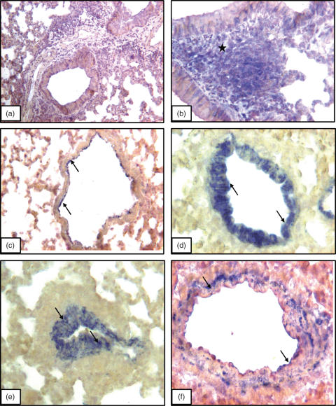
Fig. 4.
VAP-1 immunostaining: Lung section incubated without primary antibody but with secondary antibody (a) show no staining while CD3 antibody stains (asterisks) peribronchial lymphocytes (b). VAP-1 staining (arrows) in lung section from a normal A/J mouse (c) and those challenged for (d) 4 weeks and (e) 8 weeks. (f) Lung section from a BALB/c mouse challenged for 8 weeks and shows VAP-1 staining in the wall of pulmonary artery. Original magnification: a ×10; b, c and e ×20; d and f ×40

