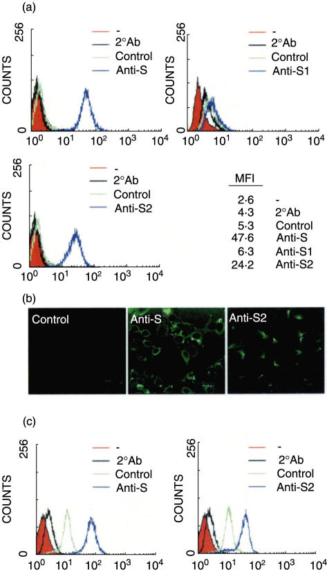
Fig. 4.
The epithelial cell cross-reactivity of anti-spike Abs. A549 cells (a, b) and HL cells (c) were incubated with a 1 : 50 dilution of anti-S, S1, or S2 mouse hyperimmune sera, followed by FITC-conjugated anti-mouse IgG, and then analysed by flow cytometry (a, c) or viewed with confocal microscopy (b). The normal mouse sera were used as the negative control. MFI: mean fluorescence intensity.

