
Figure 4.
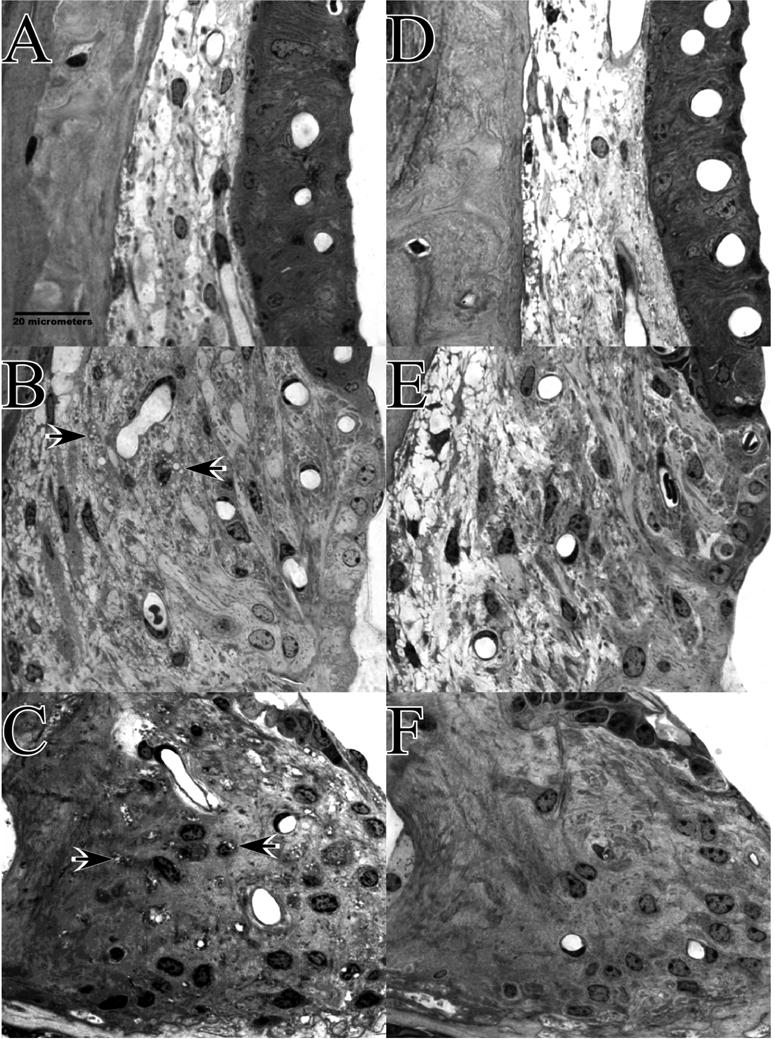
Appearance of cochlear upper basal turn lateral wall and spiral limbus in example B6.CAST mice 1–3 hrs after noise (A–C), or 8 wks after noise (D–F). A. Acutely, stria vascularis and adjacent spiral ligament appear normal. B. Some Type II fibrocytes in spiral ligament contain vacuoles (arrows) but to a much more limited extent than in CBAs. C. Spiral limbus shows vacuolization of fibrocytes in the central zone (arrows). Like the shrinkage shown in Figure 3C, this was seen in both CBA and B6 and was present to some extent in control mice. D. At 8 wks stria and adjacent ligament appear normal. E. Type II region of spiral ligament appears generally normal. F. Spiral limbus shows no significant loss of fibrocytes.