
Figure 7.
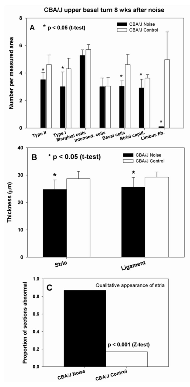
Quantitative analysis of stria vascularis, spiral ligament, and spiral limbus in cochlear upper basal turn of CBA/J mice 8 wks after noise exposure. A. Versus non-exposed controls, noise exposed mice showed significant loss of Type I and II fibrocytes, strial basal cells, strial capillaries, and (nearly complete) loss of limbus fibrocytes. B. Stria and ligament were significantly thinner than in non-exposed controls. C. The incidence of strial abnormalities was significantly greater in noise exposed mice, increasing more than 4-fold.