
Figure 8.
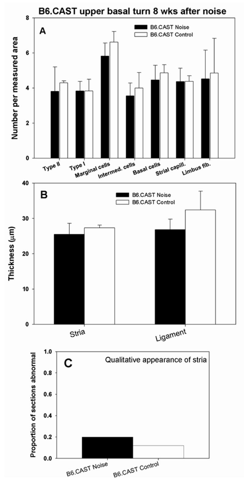
Quantitative analysis of stria vascularis, spiral ligament, and spiral limbus in cochlear upper base of B6.CAST mice 8 wks after noise exposure. No anomalies were significant versus non-exposed controls (Compare with Figure 7.) Significant thinning of spiral ligament cannot be ruled out due to scatter of data (B).