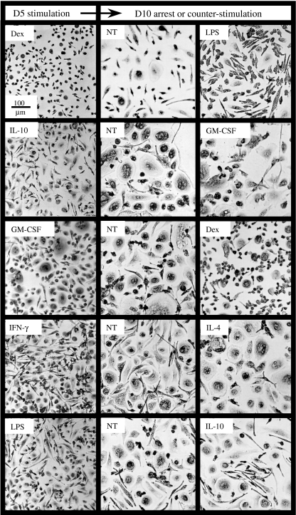
Fig. 4.
Plasticity of macrophage morphology. Mature macrophages were treated for 5 days by different pro- or anti-inflammatory stimuli (first round, left-hand panels). Cells from one well per condition were then stained with May–Grünwald–Giemsa stain. The remaining wells were then washed and cultured for another 5 days in medium alone (central panels, NT) or treated with molecules different from that used during the first round (right-hand panels). Cells were then stained with May–Grünwald–Giemsa stain.

