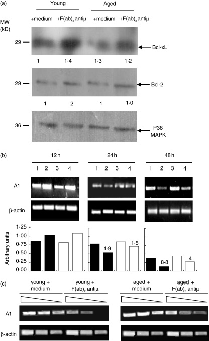
Fig. 2.
Expression of Bcl-2, Bcl-xL and A1. (a) Purified B cells from young and aged mice were cultured with medium alone or F(ab)2 anti-µ (10 µg/ml) during 24 h. The expression of Bcl-2 and Bcl-xL was determined by Western blot. p38 MAPK expression was used to determine parity of loading. The densitometric values of Bcl-2 and Bcl-xl expression are shown in relative arbitrary units at the bottom of the figure. (b) Purified B cells from young and aged mice were cultured with medium alone or F(ab)2 anti-µ for 12, 24 and 48 h. RT-PCR was performed to detect A1transcripts. Number 1 and 3 represent B cells from young and aged mice, respectively, incubated with media alone, number 2 and 4 represent B cells from young and aged mice, respectively, stimulated with F(ab)2 anti-µ. The densitometric profile of A1 expression is shown in relative arbitrary units. (c) Purified B cells from young and aged mice were cultured with medium alone or F(ab)2 anti-µ for 48 h. Semi-quantitative PCR was performed to detect A1transcripts in serial dilutions of cDNA samples. In (b) and (c), β-actin was used as internal control of RNA integrity and equal loading. Immunoblotting for A1 could not be done due to the poor quality of commercially available Ab reagents. In (a–c) one typical experiment from the three performed is shown.

