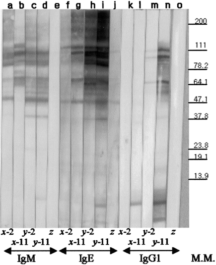
Fig. 3.
Immunoblot analysis with specific antibody isotypes of IgM, IgG1 and IgE. Antigens recognized by the specific antibody isotypes IgM (lanes a–e), IgE (lanes f–j) or IgG1 (lanes k–o) were analysed by Western blot. Sera were from two individual rats infected either with 5-L3 (x) or 20-L3 (y) or from negative control rat (z) either at 2 week ppi (lanes a, c, f, h, k, m) or at 2 week pri (11 week ppi, lanes b, d, g, i, l, n). The molecular marker is shown on the right.
The avidities of specific antibody isotypes IgM, IgG1 and IgE were analysed using sera from 1 to 4 weeks ppi and from 1 to 4 weeks pri. The avidity at RI tended to exceed that at PI for IgM, IgG1, and IgE (Figs 4a–c). In the case of specific IgE avidity, no noticeable differences were found 1, 2, or 3 weeks pri (Fig. 4c). This similarity in specific IgE avidities was also observed using another rat sera preparation (data not shown).

