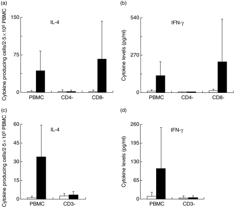
Fig. 5.
Phenotypic characterization of Ni-specific IL-4 and IFN-γ producing cells. Ni-PBMC were depleted of CD3+, CD4+ or CD8+ cells and (a, n = 6; c, n = 5) IL-4 and (b, n = 6; d, n = 4) IFN-γ production was determined by ELISpot (a, c) and ELISA (b, d). PBMC and depleted cell fractions were incubated at a concentration of 2·5 × 106 cells/ml in the absence or presence of 50 µM NiCl2. Cytokines produced spontaneously (□) and in response to Ni (▪) are depicted.

