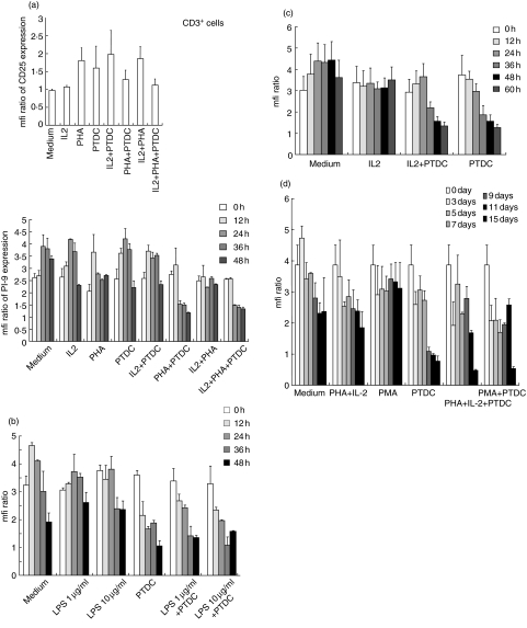
Fig. 3.
Proteinase inhibitor 9 (PI-9) expression in lymphocyte subsets following in vitro stimulation. Lymphocyte subsets were isolated by magnetic sorting, leading to a >90% pure suspension of cells that were CD3+ (T cells), CD20+ (B cells) or CD56+ [natural killer (NK) cells]. Purity was confirmed by flow cytometry (data not shown). Mean and standard deviation of triplicate experiments are given. (a) CD3 positive cells were analysed. First, stimulation was assessed by CD25 staining. Up-regulation of CD25 within 48 h was seen upon stimulation with phytohaemagglutinin (PHA) (20 µg/ml) alone or in combination with interleukin (IL)-2 (200 U/ml). Secondly, PI-9 expression was analysed in the cells. However, no stimulus-dependent up-regulation of PI-9 was seen. Pyrrolidin dithiocarbamate (PDTC) (0·3 µM) had inconsistent effects on stimulation; however, in isolated T lymphocytes, it led to a marked down-regulation of PI-9. (b) To stimulate CD20+ cells (B cells) in vitro, lipopolysaccharide (LPS) (1 µg/ml and 10 µg/ml) was applied. No up-regulation of PI-9 was evident. PDTC (0·3 µM) led to a marked down-regulation of PI-9, both in the presence or absence of lipopolysaccharide (LPS). (c) CD56+ cells (NK cells) were stimulated with high-dose IL-2 (200 U/ml) to induce rapid activation of NK cells. This did not up-regulate PI-9. PDTC (0·3 µM) led to a marked down-regulation of PI-9 both in the presence or absence of IL-2. (d) Non-adherent peripheral blood mononuclear cells (PBMC) (lymphocytes) were stimulated over 15 days upon incubation with PHA/IL-2 (PHA 20 µg/ml; IL-2200 U/ml) or phorbol-myristate-acetate (PMA) (20 ng/ml), and in the presence of PDTC (0·3 µM). Cells were collected at the given time-points and frozen until they were analysed. We saw no up-regulation of PI-9 as shown by flow cytometry. Once more, PDTC (0·3 µM) led to a marked down-regulation of PI-9 both in the presence or absence of stimuli.

