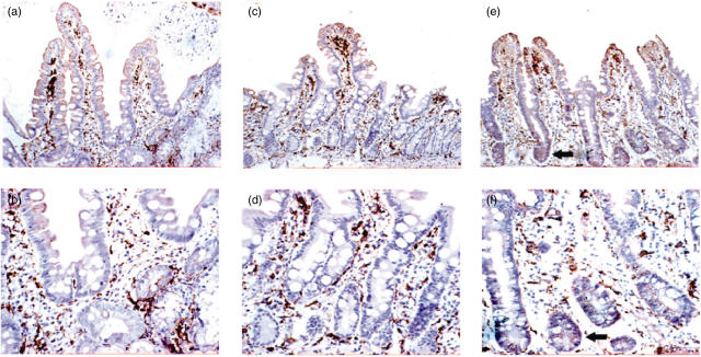
Fig. 1.
Human leucocyte antigen D-related (HLA-DR) staining of duodenal histological sections before oats challenge (a, b), after oats challenge (c, d) and after gluten challenge (e, f). The magnification of (a), (c) and (e) is ×20, with the corresponding sections shown at ×40 in (b), (d) and (f). In the pre- and post-oats sections, staining of lamina propria cells and villous enterocytes is observed. In the post-gluten sections, in addition to the above structures, staining of crypt enterocytes is observed (arrows).

