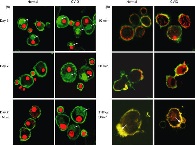
Fig 4.
Confocal microscopy imaging of major histocompatibility complex (MHC) Class II DR expression in normal and common variable immunodeficiency (CVID) dendritic cells. (a) Normal and CVID dendritic cells on days 6 and 7 of culture without treatment and 24 h after tumour necrosis factor (TNF-α) stimulation, and incubated with lipopolysaccharide (LPS)-treated CD14+ depleted peripheral blood mononuclear cells (PBMCs), were fixed, permeabilized and stained with anti-MHC class II DR fluorescein isothiocyanate (FITC)-conjugated antibody and propidium iodide. Transverse plane images show external and internal (arrow) stores of MHC molecules (green) before and after maturation. Data representative of eight separate experiments. (b) Internalization of cross-linked MHC class II DR antibody after 10 and 30 min in mature and in TNF-α-treated dendritic cells (30 min). Live cells were incubated with unconjugated mouse antibodies to MHC class II DR on ice and cross-linked with goat anti-mouse Alexifluor 586-conjugated antibodies (red) at 37°C. Cells were subsequently fixed and stained with FITC-conjugated cholera toxin (green). Coincidence of MHC class II DR and GM1 proteins is seen as yellow, accumulating at contact points (arrow) between monocyte-derived dendritic cells (MdDC) and LPS-treated lymphocytes. Cells representative of five experiments.

